切片显微镜-Slicing microscope
显微镜是人类20世纪最伟大的发明物之一,它把一个全新的世界展现在人类的视野里,人们借助它可以看到微小动物、植物纤维等各种东西的内部构造,有助于医生识别病毒治疗相关疾病。传统的显微镜只能用肉眼查看局部的图像信息,无法将图像信息进行保存和分享。
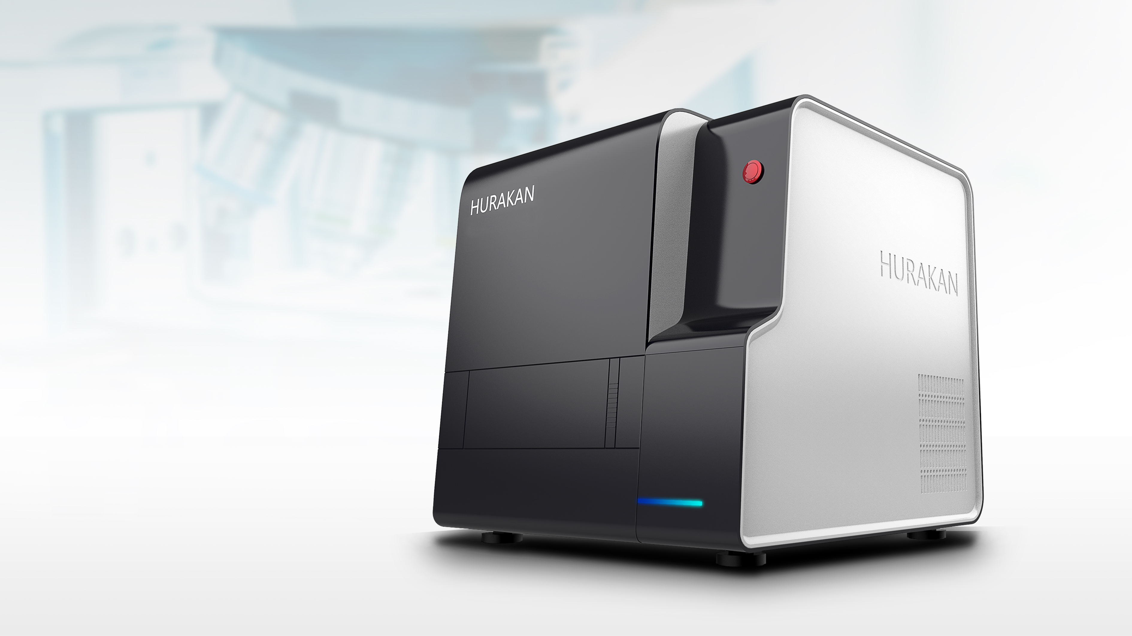
HURAKAN联合客户共同开发数字化的切片显微镜,通过高分辨率的摄像头将切片样品高清放大,同时直接转化成高清数据图像,便于保存和分享图像,根据患者的切片样本分析,制定最适合患者的治疗方案。
Microscope is one of the greatest inventions of human beings in the 20th century. It shows a whole new world in the human field of vision. People can see the internal structure of tiny animals, plant fibers and other things with the help of microscope, which is helpful for doctors to identify viruses and treat related diseases. Traditional microscopes can only view local image information with the naked eye, which cannot be saved and Shared.
HURAKAN has cooperated with customers to develop a digital microscope for slicing, which can be directly converted into high-definition data images through high-resolution cameras to facilitate the storage and sharing of images. Based on the analysis of slicing samples, the most suitable treatment scheme for patients is formulated.